
The good news is that a lump doesn’t always mean bad news, and even if it is a type of cancer there may be a number of treatment options available.
Here we'll look into how we go about determining what the lump is and what possible treatments options are available.
Breaking down the jargon
Terminology can be confusing and you may hear the terms lump, mass, growth, tumour, neoplasia or cancer being used interchangeably. Broadly speaking lump and mass are descriptive and used when we don’t yet know what we are dealing with.
A lump may be due to inflammation, such as around a hair follicle or foreign body such as a thorn, rather than due to a growth. A growth/mass/tumour/cancer implies a form of cancer and occurs when the normal process controlling cell growth, division and repair is disrupted and becomes uncontrolled. However, this growth/mass/tumour/cancer can either be benign, meaning that it behaves in a non-aggressive way and does not spread elsewhere within the body, or malignant meaning that spreading to other areas in the body or local invasion of surrounding structures, is likely to occur.
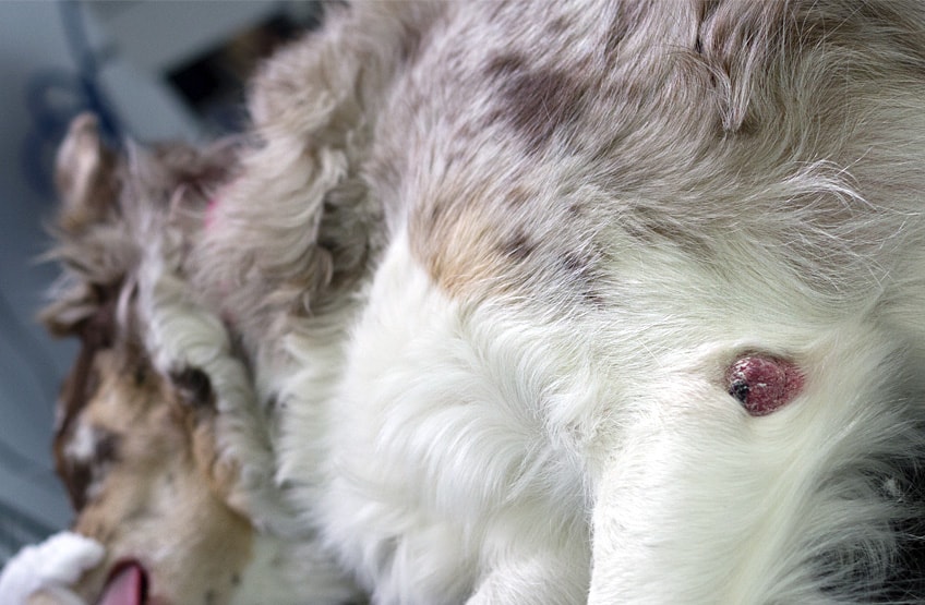
So what are we dealing with?
The first thing the vet will need to do is assess the lump. We may check for:
- Is the lump firm or soft?
- Is it painful?
- Is it mobile and ‘free’ within the skin layer or attached to deeper structures?
- Has the hair over the top of the lump been lost (if located on haired skin) and has it become red, sore or ulcerated?
Although sometimes it may seem obvious that the mass is a fatty lump, wart or cystic lesion, some masses may be surrounded by fat or mimic other masses. Therefore, to be certain what we are dealing with the lump should be sampled.
Contact us if your pet has a lump
Why sampling is so useful?
The aim of sampling is to determine the nature of the lump which then guides the vets approach of treating it. The questions we are trying to answer when taking a sample are;
- Is it inflammation or a tumour?
- If it is a tumour what type is it and is it benign or malignant?
- Is monitoring appropriate?
Planning - can the lump undergo local excision (removal with only a small amount of the surrounding normal tissue) or does it require more advanced surgical techniques to take away larger amounts of surrounding tissue to prevent recurrence?
What type of sample will the vet take?
This will depend on a number of factors such as the location of the mass, it’s size, the temperament of your pet, the location of the mass on the body, suitability for sedation or anaesthesia and associated costs.
All of these factors will be discussed with you, however if you are unsure or have any questions or concerns please contact us.
Broadly, there are two sampling techniques. The first is known as cytology, using a technique know as a ‘fine needle aspirate’ or FNA. A little needle, like a blood sample needle, is inserted into the mass. Suction is applied with a syringe. The needle is removed and the contents of the needle hub expressed onto a microscope slide. This is then either assessed in-house or sent to an external lab for an expert opinion from a pathologist. This technique is minimally invasive, it can usually be performed straight away in your consultation or you may be booked in for the sample to be collected in a longer consult the following day.
It often doesn’t require any sedation/anaesthetic if your pet is tolerant. It is relatively inexpensive compared to biopsy and results are usually known within a couple of days. Although most the time the lab will be able to give us an answer, as this techniques involves collection of a few cells only and not a chunk of tissue, there is always a possibility that we don’t get a conclusive answer and have to consider ‘plan b’ (a biopsy). The other technique is to take a biopsy. This involves taking a chunk of tissue to send to the lab for histopathology. This is a surgical procedure and so involves admitting your pet for the day for the procedure to be performed under general anaesthesia. The advantage is that it gives the lab more tissue to work with so there is less chance of an inconclusive result. However it does involve an anaesthetic and is therefore more expensive. Usually the lump is not completely removed by biopsy, so your pet would require a second surgery for this.
Can't we just remove the mass?
Accepted veterinary gold standard involves sampling of the lump first. It allows the vet to know what the lump is and therefore choose the optimum treatment path and how big a margin of normal tissue should be removed. It removes some uncertainty and the risk of recurrence and complications. In some circumstances, however, compromises have to be made, so we recommend you discuss options with your vet to create an individual treatment plan that suits you and your pet. On occasions, if the mass is very small and located in an area of the body where there is plenty of ‘spare’ skin, biopsy may result in complete excision. However, as previously mentioned in some circumstances the histopathology result may indicate that the mass has spread into the surrounding tissues and so a repeat surgery may be required to remove more skin.
If in doubt, contact us!
It is difficult to tell from examination alone what the lump is as there are many different types of lumps. Sampling allows us to know what the lump is and more about how it is likely to behave, such as whether it is likely to grow or spread. This way we know whether we can stop worrying about the lump or if we need create a treatment plan.
Having pet insurance is important for when matters arise such as finding a lump on your pet. Find out more about pet insurance advice here.
If you find a lump on your pet, call your local Calder Vets branch to book an appointment so that we can carry out the necessary checks.
Our Services