
Our veterinary surgeons at Calder Vets use many modes of imaging, from radiographs (x-ray) to computed tomography (CT) scan. Each type uses different methods to create images, and each is best suited to different body parts.
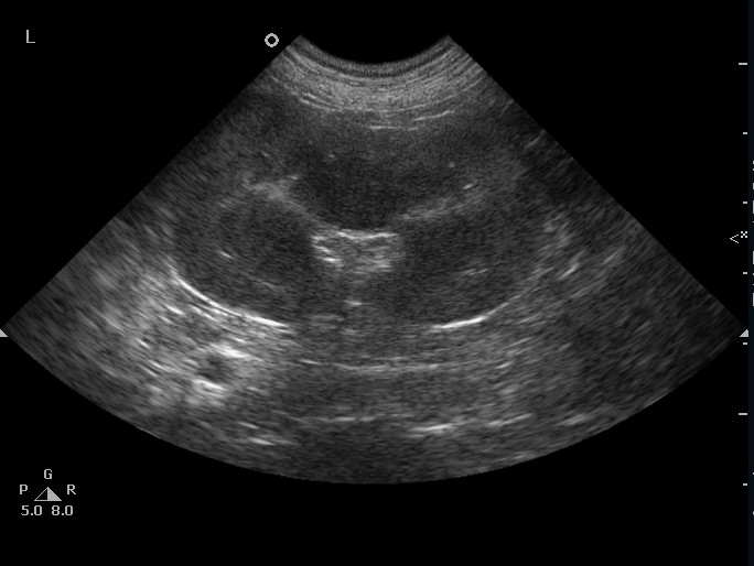
Ultrasound machines employ the same technology used to generate pictures (sonograms) during human pregnancy, transmitting waves into the body to create an image. This can be a really useful tool to image structures, allowing vets to look at the architecture of an organ or cavity. For example, when one of our patients has a tumor or ingest something they shouldn’t have, an ultrasound can be helpful to locate these objects and characterise them before starting treatment. Ultrasonography can be lifesaving in an emergency situation.
Ultrasound is a non-invasive procedure that has no side effects, although occasionally sedation or general anesthesia is needed to help nervous patients remain still to allow for high-definition and clear images to be obtained.
Apart from abdominal scanning, ultrasound also allows us to study one of the most complex parts of the body, the heart. Ultrasound is one way we can evaluate the function of the heart and diagnose specific heart diseases. This is called echocardiography, which simply means an ultrasound of the heart. Your vet will evaluate how the blood is flowing through your pets heart as well as whether the heart valves are functioning properly during a heartbeat.
After having an ultrasound your vet may recommend using computed tomography (CT) and magnetic resonance imaging (MRI) to investigate certain heath issues further. This is to gain as much information as possible about your pet’s condition to allow for the best treatment plan possible.
Our Services